The Other Bacteria...
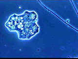
Amoebae...
What does it mean when I see an increase in Amoebae in my system ? It depends upon what the rest of the biomass looks
like. If the floc is small, weak, dispersed, you may have a very young sludge age. Typically the presence of amoebae
indicates a high loading of food vs. the amount of biomass available to eat the organics. It may mean the sludge is
young, or if you have had rotifers in the past and were old, it may mean a recent high loading of BOD that is forcing
the sludge age to a younger age. Usually you can expect high solids in the effluent and higher BOD levels if amoebae are
present in significant numbers.
Daily microscopic analyses is helpful in documenting where you are today, where you have changed since the previous day
and how to react to changes proactively as opposed to when they have become critical !
What should I do if there is a significant change in my higher life forms and all of a sudden there is an increase in
Amoebae ? First check to see why they have increased ? Is there a change in loading that might impact other areas ?
Check your nutrients in this case if applicable to your plant. The biggest mistake people make when a high loading comes
through or a spill, especially at industrial plants is not to increase nutrient levels when high loading occurs. You might
want to adjust your wasting or RAS levels. Some plants add bioaugmentation products in cases of higher loadings. You might
need to slightly increase the dosage of product. If using micronutrients, adjust these levels also if the loading is
significant. You might need to check the Bed levels in your clarifier. Check your TSS off your clarifiers.
All amoebae presented in this class are parasitic. There are many others that are free-living. The typical life cycle
involves infection of the host with the trophozoite multiplying by binary fission and in some cases, producing cysts.
Diagnosis in all species requires microscopic evaluation of either trophozoites or cysts. ( Not all species of amoebae
have a cyst stage ).
Entamoeba histolytica : Agent of intestinal amebiasis. This is a pathogenic organism commonly recovered from man
but has also been reported from lower primates, dogs, and cats. The typical location is the cecum, colon or rectum with
extra-intestinal locations common. E. hostolytica trophozoites damage the intestine by attaching to and lysing host cells
and secreting enzymes that disrupt intercellular connections. The presence of some bacteria and deficient protein intake
may contribute to parasite virulence. Erosion or ulceration of the colon mucosa is common with classical "flask-shaped"
ulcers. These are the result of trophozoite undermining the mucosa and submucosa. Clinically, diarrhea with dysentery
(diarrhea with abdominal pain, straining and blood in the stool) is the main problem in intestinal infections. Clinical
problems associated with extraintestinal lesions depend on the organs involved.
Although a problem in humans, E. histolytica rarely causes clinical problems in other non-primate animals. Although dogs
and cats are susceptible to infection, it is not likely that they contribute to the epidemiology of human infections and
are most likely infected themselves from exposure to infected human feces. Treatment should be used to destroy
trophozoites, relieve symptoms and control secondary infection. The drug(s) of choice are metronidizole and
diiodohydroxyquin. Tetracycline therapy is used in combination with both drugs to combat secondary bacterial infections.
Hepatic amoebiasis usually responds to metronidazole therapy but may not be totally effective. Chloroquine therapy is
often used when contraindications occur but has no effect on intestinal trophozoites. Both trophozoite and cyst stages
occur. Man is also infected with a variety of other non-pathogenic amoebae that require differentiation from
E. histolytica.
Acanthamoeba spp. : Infections of this type are becoming more common in both humans an canines. This organisms are
ubiquitous free-living amoebae that under specific circumstances can cause infection and disease in some animals. Acanthamoeba infections in canines are rare but reported. Young dogs appear to be the most susceptible and infections in greyhounds, German shepherds and an Akita are recorded. Clinical signs include mild oculonasal discharge, anorexia and lethargy. Increased body temperature, respiratory distress and occasional neuralgic signs. Laboratory findings are generally
nonspecific. Premortum diagnosis is rare. New cases of Acanthamoeba respond to sulfonamides while established cases
are generally treated with amphotericin B. Corneal ulcers resulting from infections with Acanthamoeba infections in
contact lens wearers appear refractory to treatment. The life cycle of Acanthamoeba spp appears similar to that of
Naegleria fowleri and several cases of chronic PAM have been attributed to this organism.
Entamoeba coli : This amoebae is similar to E. histolytica but is not considered a true tissue invader and thus is
non-pathogenic. Differentiation from E. histolytica is based on morphologic differences and the number of nuclei (4 in E.
histolytica, 8 in E coli) in the cysts and nuclear morphology in the trophozoites.
Naegleria fowleri : Agent of primary amoebic meningoencephalitis (PAM). This is a free-living amoebae that
occasionally infects humans with a high case fatality ratio. It was first identified as a problem in 1965 and over
150 cases have now been recorded. Most human victims have a history of exposure to warm, fresh or brackish water, such
as swimming pools, ponds, lakes or streams. Transmission to humans appears to occur when the nasopharyngeal mucosa is
invaded by the flagellated trophozoite form of the organism. These trophozoites migrate through the nervous system to
the brain where inflammation occurs followed by death. Most cases are diagnosed at autopsy by microscopic identification
of the motile amoebic trophozoite form in cerebrospinal fluid. The clinical course of the disease is generally 5 to 7 days
after exposure. No effective treatment exists but favorable response to intravenous administration of amphotericin B has
been reported. Although a variety of animals are known to be susceptible to infection, little is known about the prevalence
of this organism in domestic or wild animals.
Pics of Amoebae...