Lagoon Microorganisms...
Algae...
What are algae ? : The term "algae" is used for some lower plants and many, often unrelated groups of
microorganisms that are able to perform photosynthesis. Photosynthesis (converting light energy into chemical energy)
is performed in parts of the cell called chloroplasts. They can be found in different shapes and colours and in many different organisms. Not all these organisms are green. Diatoms, Chrysophytes and dinoflagellates have yellow to brown chloroplasts. There are brown algae (Phaeophyta), red algae (Rhodophyta) and many other groups of unicellular algae
in many shades of green. The blue green Cyanobacteria also photosynthesize.
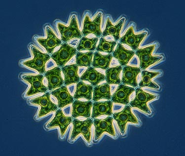
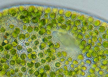
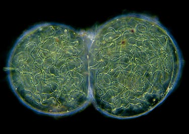
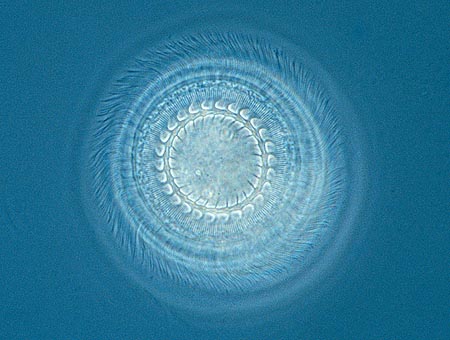
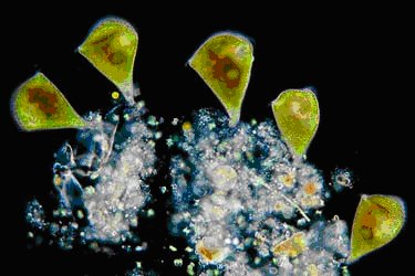
A very diverse groups of freshwater algae are the Chlorophytes or Green algae. Based on the compounds of the
photosynthetic pigments and several other characteristics they seem closest related to plants. A common green algae is Hydrodictyon, the water net. It is a related to Pediastrum (top image) But it forms a bag-shaped colony. Like Pediastrum
each individual cell can develop into a new colony. You can imagine that since the colony contains thousands of cells Hydrodictyon can reproduce very rapidly. And unlike Pediastrum, Hydrodictyon can grow large, almost 30 cm. in length.
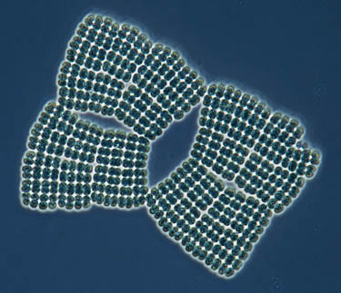
Blooms of Hydrodictyon can be a real problem for water treatment plants. The image shows a part of a small colony (left)
and three individual cells of a big colony. Inside each of these cells a new colony can be formed.
If you have a birdbath with water that has turned red you may find the green algae Haematococcus pluvialis. This species illustrates that green algae don't always have to look like green algae. The chloroplast often turns red when conditions become unfavourable. Haematococcus swims with the aid of two long flagella. Many species are flagellated and motile, other are immobile but often have a flagellated stage at some time in their life cycle. Certainly one of the most spectacular flagellated green algae is Volvox. It forms a spherical colony. All the small cells of the colony possess two flagella and
a small eyespot. With this the colony is able to swim towards the light. Volvox has interesting ways of reproduction.
Find out much more about it in the Micscape article Volvox, one of the seven wonders of the microworld. A small rather inconspicuous green algae without undulipodia is Chlorella. It can be found as an endosymbiont inside ciliates, hydra
and other animals. They raise Chlorella as if they grow crops in a greenhouse.
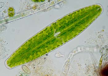
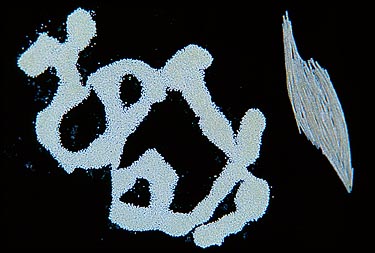
Many green algae form long filaments. The cells stay attached after they divide. Some genera, like Spirogyra, Mougeotia
and Zygnema can become so numerous they form dense mats of growth in surfaces of ponds, so-called pond scum. This pond
scum is interesting for a study under the microscope. If you squeeze it in a jar you will collect many interesting
organisms. And the filamentous algae are at least as interesting. The chloroplasts have distinct shapes. In Spirogyra the chloroplast runs through the cell like a helix. The image also shows the nucleus hanging on fine threads. Spirogyra and related algae like the Desmids are conjugating green algae. These desmids are so beautiful they deserve a page for themselves.
Bacteria...
Bacteria are the most ancient life forms Most bacteria are so small that under a light microscope you can only see them as little dots. Some groups however grow to larger sizes and have spectacular shapes.
The top picture shows an example of the beauty of one of the more advanced groups of bacteria, the cyanobacteria, or blue green algae. For a long time they were regarded algae since they performed photosynthesis. But they are nowadays regarded bacteria: they lack a membrane bound nucleus. DNA is present in a loop in the cytoplasm.
Because they have a very distinct blue green colour a bloom of cyanobacteria is clearly visible when a pond or ditch becomes blue-ish green.
Bacteria were the most prominent creatures in the early stages of life's history almost 4000 million years until 600 million years ago. Fossils called stromatolites can still be found and were made by Cyanobacteria.
Cyanobacteria were essential for the developing of more complicated life forms They produced the oxygen in our atmosphere. This oxygen is needed for the much more energy efficient metabolism of mufti cellular organisms. Moreover, the photosynthetic abilities of bacteria were presumably incorporated by the more advanced organisms. At present bacteria have an essential role in the recycling of matter. Bacteria are everywhere. Inside your mouth live more bacteria than the number of people who have ever lived.
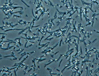
The little animation on the left shows spirochaete bacteria. They are fast movers that have many flagella that unlike other bacteria lay inside the cell. Many other bacteria use flagella (whip-like structures) for locomotion. The flagella of bacteria are quite interesting since they have a simple but very ingenious design. A bacterial flagellum is a miniature mechanical device with a movement that is caused by rotation of the shaft, the part where the flagellum is attached to the cell. It is an extra cellular structure that works like a little propellor.
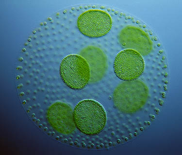
Nostoc is a cyanobacteria that forms large spherical colonies. They are common and can just be seen with the naked eye. Most bacteria are only visible with the strongest lenses of the microscope. An easy way to collect them (and also many small protists) is by placing a cover slip on the surface of pond water. After a night whole colonies of bacteria will grow on the underside of the cover slip. Carefully placed on a slide (an extra drop of water may be added) the growth under the cover slip can be observed with the 40X or 100 X objective to see the finest details.
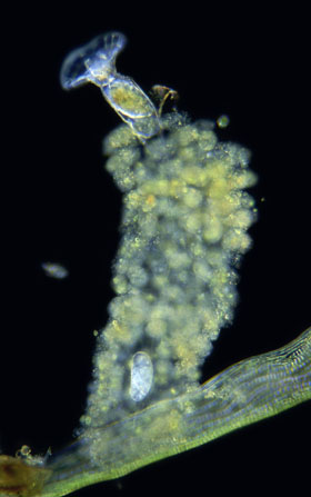
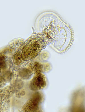
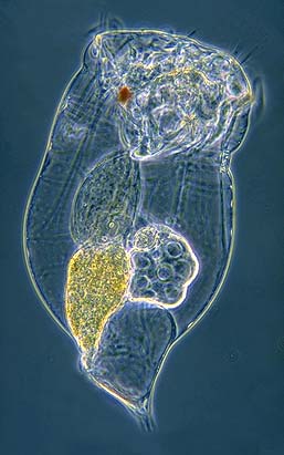
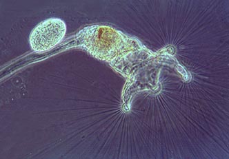
Rotifers...
Rotifers are multicelled animals. Because they are so small most people have never heard of their existence. They are
about the same size as the larger unicellular organisms. They don't have a lot of cells, less than 1,000, but they have
some very special attributes. They are wonders of miniature design. At the front of the body they possess a crown of hair-like cilia. They locomote by using the crown of cilia (the corona) to propel themselves. Some species walk with
head and foot. Their foot can secrete a sticky substance that enables them to attach to a surface. They also use the
crown of cilia to wave food into their mouth. There the food is passed into the 'mastax' where two so-called "trophi"
process the food before it is directed towards the gut. Rotifers are so transparent that all these organs can be observed easily. They have one or two light sensitive red eye spots.
There is an enormous variety of spectacular body shapes, all to suit the different lifestyles or environmental conditions.
Because many species make so-called resting spores which are easily carried by the wind, they can be found anywhere if
there is a little bit of water. Even in a roof gutter or in birdbaths. The corona, the crown of cilia, can vary in design. Some species don't use it for locomotion but have developed very special capturing devices. The rotifers from the genus Collotheca live attached to a substrate and collect tiny microbes like bacteria with extremely elongated cilia. To see
these anglers under the microscope is a beautiful sight.
Amoebas...
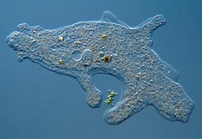
What are Amoebas ? : Amoebas (Phylum Rhizopoda) are unicellular protists that are able to change their shape constantly. Each species has its own distinct repertoir of shapes.
How does an amoeba locomote ? : Amoebas locomote by ways of cytoplasmic movement. (cytoplasm is the cell content around the nucleus of the cell) The amoeba forms pseudopods (false feet) with which they "flow" over a surface. The cytoplasma not only flows it also changes from a fluid into a solid state. These pseudopods are also used to capture
prey, They simply engulf the food. They can detect the kind of prey and use different "engulfing tactics". The image
shows several cell organelles. Left from the center we can see a spherical water expelling vesicle and just right of it,
the single nucleus of this species can be seen. Other species may have many nuclei. The cell is full of brown food
vacuoles and also contains small crystals.
Amoebas can grow to exceptionally large sizes for a unicellular organism. Some species are 5 millimetre long. They
can reach such a size by having many nuclei within their single cell. Find out much more about Amoeba in the Micscape
article Amoebas are more than just "blobs". Amoebas are not known to have any sexual way of reproduction. They only
multiply by cell division.
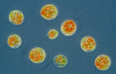
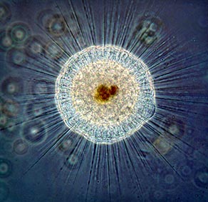
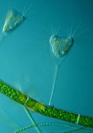
Sun animalcules : or Heliozoa, phylum Actinopoda (=ray footed) have axopodia, long stiff projections of cytoplasm. They cannot locomote that much with them but they are excellent for capturing prey. The prey, usually small protists,
sticks to the axopods and will be covered by the cytoplasmic flow running along the axopods and transported towards the
cell Sometimes Heliozoa can be found feeding on the same prey. In this image 2 individuals of the species Actinophrys sol
are feasting on a diatom.
Some amoebas form a shell from found material that they glue together to form a protective covering. Often sand grains or other particles are used to make the construction, called a "test" stronger. You can see how this shelled amoeba hangs in
its test keeping contact using thin pseudopods. Long pseudopods are extruded to capture prey.
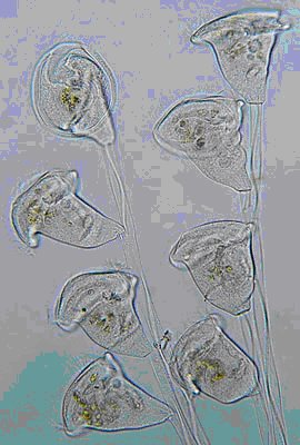
Ciliates...
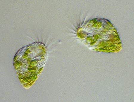
What are Ciliates ? : Ciliates are unicellular protists that can be recognised by their hairlike "cilia". They use them for locomotion and for feeding. Some ciliates are very small, not much larger than the largest bacteria. Others like
the "trumpet animalcule" Stentor can reach a size of two millimetres so it can be seen with the naked eye. Paramecium does not become much larger than 0.3 mm.
Abowe the centre of the organism you see the feeding opening. The cilia make a current to sweep in bacteria and other food particles. At the base of the feeding opening the food has been enclosed by a vacuole. Food vacuoles are used to transport the food through the cell. Star-shaped contractile vacuoles or water expelling vesicles are used to balance the amount of water in the cell. Two nuclei bear the genetic information.
Why cilia ? : When you are less than a millimeter in body size, water acts like a thick syrup so swimming like a
fish would not be an efficient method of locomotion. If you want to swim fast and with manouverability you need cilia,
tiny hairs acting like paddles. Most ciliates are superb swimmers, some species are so fast that they are hardly visible
when observed.
In multicellular organisms the many cells are specialised (differentiated). Different types of cells are able to perform
all kinds of tasks (i.e. act as nerve cells, muscle cells, blood cells etc.). Unicellular organisms possess organelles, special structures inside or on the cell that help to perform all kinds of tasks. Food vacuoles and the water expelling vesicles are such organelles. Many ciliates have developed all kinds of very special organelles. Paramecium uses so-called trichocysts; tiny pointed filaments that can be fired at a predator when threatened.
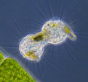
Myonemes : They are fibers that act like muscles. We can see them in the stalks of bell animalcules like
Vorticella and Carchesium. They are able to contract the stalk with exceptional speed. In the righthand image the
myonemes are visible as the light wavy bands inside the stalks. In many ciliates fused cilia can be seen. Groups of
cilia are fused into sheet-like membranelles. As undulating sheets they sweep in food particles. They can be seen in
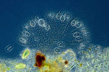
the bell animalcules. Other ciliates have thick round bundles of cilia called cirri which act like legs and enable the organism to actually walk over a surface. To coordinate these cirri independently they have nerve-like organelles called neurofibrils. The top image of Euplotes and Stylonychia shows both types of cilia.
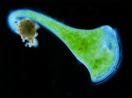
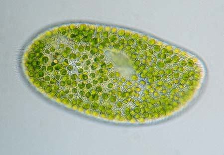
The trumpet animalcule Stentor is one of the biggest ciliates. Extended they can be 2 millimeters long and just visible
with the naked eye. These green Stentors show a clever trick several other small organisms use. They are green because
they make use of a symbiotic green algae called Chlorella. The page about Green algae will show these algae in Close up.
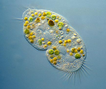
Ciliates usually multiply asexually by fission. The cell divides into two individuals. Here we can see a Paramecium undergoing fission. Once in a while we may also encounter a process which is called conjugation. These two ciliates
of the genus Spirostomum cling to each other side by side and fuse together. In a complicated process they exchange
the genetic material from their nuclei. After conjugation there is no offspring, the individuals have "renewed"
themselves instead. Most animals, even humans, have cilia similar to those of ciliates. These cilia are used inside
our body, for example, to transport food particles or other material.
Make your own hay culture : It is not so difficult to raise Paramecium or other protists with a hay culture. Just
boil a bit of dry grass for 15 or more minutes. Put the water in a bowl. Wait for a few days until a layer of bacteria
begins to develop on the surface. Then add a few specimens that you collect from pond water (a stereo microscope is always very useful for sorting them out). But you can also put the bowl of water behind a window (out of direct sunlight) and just wait for what the wind carries. Many resting spores of microorganisms are carried with the wind.
Crustaceans...
What are Crustaceans ? : This is the group of animals that is best known for the crabs and lobsters. But they have many microscopic relatives. With the insects they belong to a large group called the arthropods. They all have segmented limbs and a hardened external skeleton made of chitin.
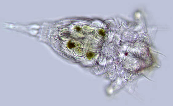
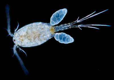
Which crustaceans can we find in fresh water ? : There are three major groups of microscopic crustaceans that you might encounter when you look at a drop of pond water: the water fleas, the ostracods and the copepods. The most familiar
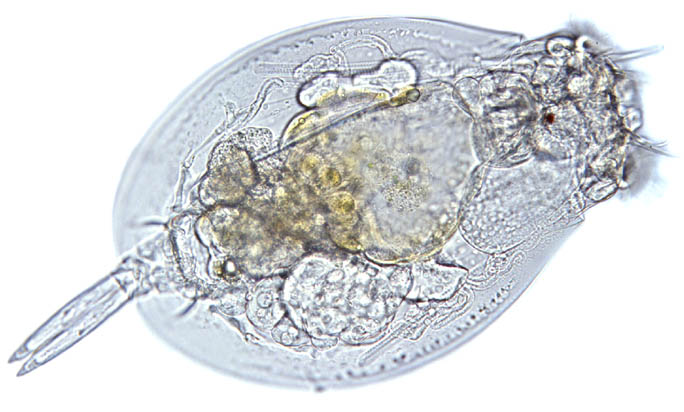
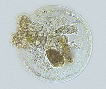
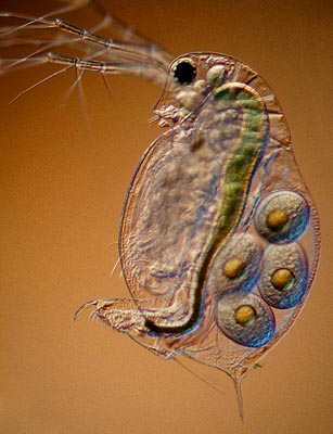
are the water fleas. They are the most numerous organisms in freshwater zooplankton. They can be seen with the naked eye because some species can reach a size of almost six millimetres. With a good hand lens you can observe many interesting features.
One of the most obvious features are the large antennae. They use them for locomotion. Above the antennae you can see the large eyes. It looks like a single eye but it consists of two compound eyes that are fused together. Inside the bivalved protective shell called the carapace lies a row of five or six pairs of feet they use to filter food (small algae) The
food can be seen as the yellow brown substance. Left of the gut, eggs can be seen. Find out much more about waterfleas
in the Micscape article Waterflea Anatomy ! The little heart is easy to observe when you examine a live specimen under
the microscope. You can count the pace of the heart beat with a pencil and a stop watch. Just tap the pencil on a piece
of paper following the pace of the water fleas heart during one minute.
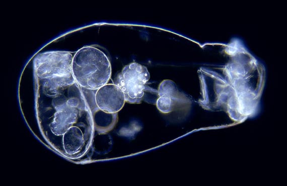
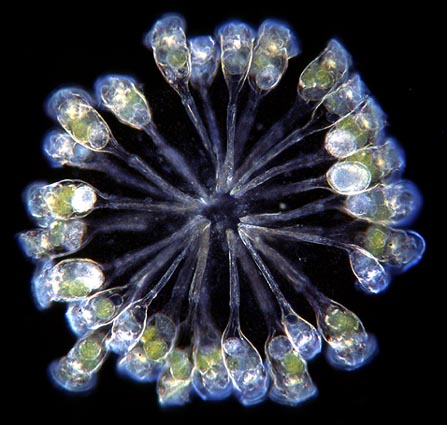
The ostracods (see top image) are very interesting little crustaceans. Their shell is even more protective than that of
the water fleas. They can shut the two halves completely and withdraw themselves in it. Their shells are so strong they fossilize well. Their fossils are very important in the study of ancient earth layers. Prehistoric earth layers can be
dated by determining the kind of species that inhabited the layer.
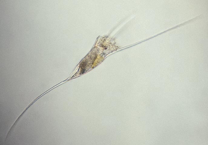
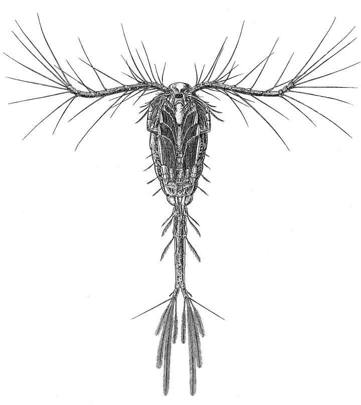
The last group of microscopic crustaceans depicted here is that of the copepods. Their body does not have a shell like appearance but is more slender and segmented. In freshwater they are not as numerous as the water fleas but in the ocean
they can be found in astronomical numbers. We may even consider them the most numerous animals on the planet. They are
the major food source for fish and even whales.
Diatoms...
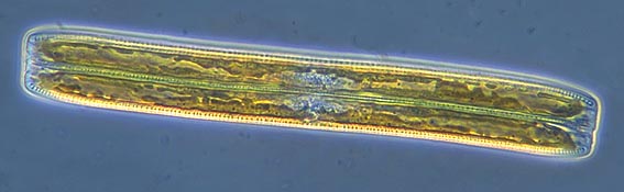
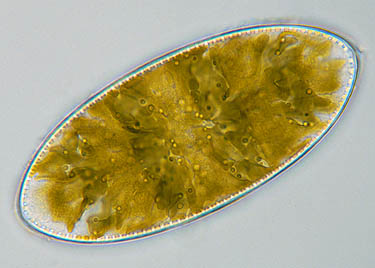
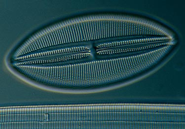
What are diatoms ? : Diatoms are delicate unicellular organisms that have a yellow-brown chloroplast that enables
them to photosynthesize. Their cell walls are made of silica almost like a glass house. The construction of the cell wall, called the frustule, consists of two valves that fit into each other like a little pill box. The colour of the chloroplast
is yellow-brown instead of the green we know of other creatures that use light as a source for energy. There are two different groups of diatoms, the pennates which are pen-shaped and the centric which are like a cylinder. In fresh water
most diatoms you will see are of the pennate type.
Where can you find diatoms ? : At the end of the winter they are most numerous in fresh water. They will cover surfaces of aquatic plants or poles and wooden borders of ponds. If you like to study them you can scrape the brown
growth with a flat piece of plastic. You can also use a sponge. For the free living (plankton) species a special fine
mesh plankton net is very useful.
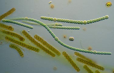
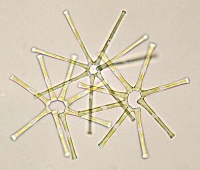
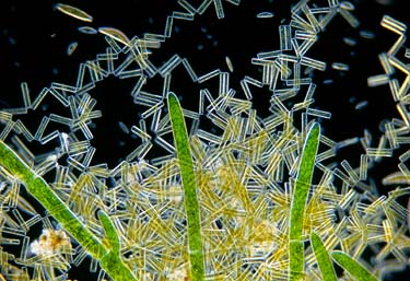
Many species of diaom stay connected after the cells divide. They form colonies, long chains. Sometimes only the tips are connected and they form a zig zag pattern. The cells of diatoms are ideal subjects for study under the microscope. They
show complex patterns of very fine punctures and they often have all kinds of ornaments. The best way to make the beautiful structures of the frustule visible is to remove the content from the cell. Hydrogen peroxide can be used for this. But it
is also possible to find empty frustules of dead diatoms. The extremely fine pores in the frustule of certain species of diatoms are used to test the resolving power of a microscope's lens.
Diatom's locomotion : Pennate diatoms, which developed later in the earth's history, are able to locomote in a
slow gliding fashion in the direction of the length of the cell. The mechanism for this is still not well understood but
it seems that through the slit alongside the cell (the raphe) tiny microfibrils protrude. With these they can move over
a surface. In the picture of the cleaned diatom above the raphe can be seen as the thin central horizontal line. There
are two different groups of diatoms, the pennates which are pen-shaped and the centric which are like a cylinder. In
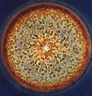
fresh water most diatoms you will see are of the pennate type. In marine waters the variety of body shapes is much
greater. In the ocean they form the main part of phytoplankton, the photosynthetic organisms that float with the
current. This image shows the top view of a large Coscinodiscus, a marine species that can just be seen with the
naked eye. Clearly visible is the fine net-like structure of the siliceous cell wall. The yellow-brown chloroplasts
used for photosynthesis are also easy to see.
Protozoa & Flagellated Protozoa...
What are Protozoa ? : The name "protozoa" is used for the more animal-like single celled organisms like amoebas
and ciliates. The term "algae" is used for the more plant-like microorganisms. But the distinction is often vague.
Dinobryon (top image) has chloroplasts for photosynthesis but it can also feed on organic matter. It is even able to swim. Such an organism can neither be animal nor plant. Nowadays, all these unicellular organisms that are neither animals,
plants, bacteria or fungi are called "protists".
What are flagella ? : A flagellum, also called undulipodium, is a whip-like structure used for locomotion, for
feeding or other purposes. Almost all organisms have flagella (at some stage in their lives). We humans have them in our bodies. Even our own spermatozoa can be regarded as flagellates. All these flagella have a similar basic design.
Only Bacteria have flagella of a totally different type. The cilia of Ciliates are just shorter versions that act
different. A cilium beats like a small pedal and generates a sideways motion. Most undulipodia act more like a
propellor, wavy motions push or pull an organism in a forward direction.
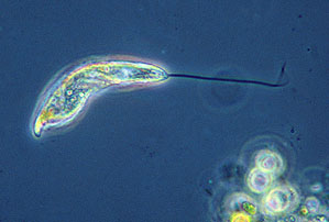
A single nucleus contains the genetic material. Bright green chloroplasts transform light into energy. The energy is stored in storage grains. A long flexible undulipodium used for locomotion. Near base of the flagellum we find a round contractile vacuole. It helps the organisms to remove surplus water. Because living tissue is saline, organisms living in fresh water take up water. A red eyespot is clearly visible. In fact the real light sensitive organ is the swelling near the base of
the flagellum. The red area makes sure only light from one direction is detected. Since the light sensitive organ is
directly connected to the flagellum, Euglena is perfectly able to swim towards a light source.
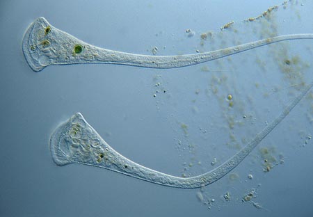
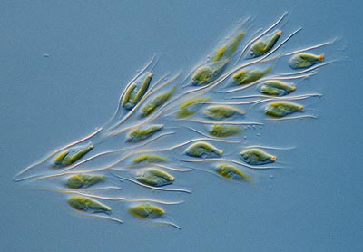
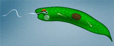
An interesting flagellated protist to study is Euglena. Some of its many features are shown here. Euglena can become so numerous it may turn the water of a pond bright green. Apart from using their undulipodium Euglenids are also able to locomote by flexing their bodies and changing their body shape, so called Euglenoid motion. Euglenids have the ability to lose their chloroplasts. If you would keep Euglenids in the dark they start feeding on organic matter and may loose their pigment. There are many species without chloroplasts.
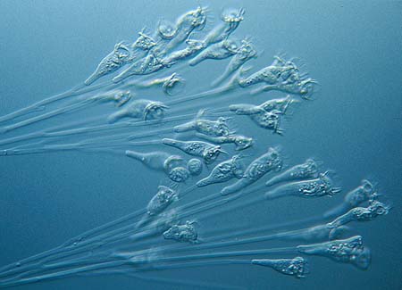
Most flagellated protists have two or even more flagella. Often we see one flagellum trailing behind like a kind of
rudder. Flagella and cilia are transparent and thin so it is not easy to see them with a microscope. Special contrast techniques like dark field illumination or phase contrast help to make these structures visible. Dinoflagellates
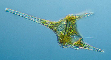
(Dinophyta) are flagellated protists. Many species have a beautiful textured armor made of cellulose plates. One of
their two flagella runs as a spiral through a groove along the cell. The other projects downwards. A spectacular species
is Ceratium hirundinella. It is not just protists that use flagella for locomotion. Even our own spermatozoa have a flagellum. Not all flagellated forms are free swimming. Many live a sessile life. Spongomonas is a colony of flagellates living in a gelatinous matrix. they use their flagella to gather food.
Make your own culture of Euglena : It is relatively easy to make a so-called "earth culture" for several kinds of photosynthetic protists and algae, including Euglena. Put a small amount of garden soil in a bowl of water and cook it
until it is sterile. Place the bowl behind a well lit window but not in direct sunlight. Now add a bit of pond water or choose an algae by using a pipette. Make sure you don't add algae eating species like water fleas. The nutrients from
the earth are enough to grow millions of euglena or other interesting species. Because Euglenids moves towards the light
you can do all kinds of experiments with them. You don't even need a microscope for that. The green swarm can be seen
with the naked eye. Perform your own Protozoan pet tricks by illuminating different sides of the bowl.
Termite gut symbionts : For those who live in the warmer areas of the world there is a very interesting place to
find truly amazing protists. Inside the gut of termites live flagellated protists that help the termites to digest wood.